
Sleeping brain: How it Dreams
- Ayesha Jannat

- Dec 24, 2022
- 2 min read
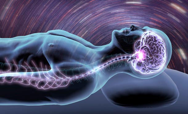
An important aspect in the explanation of dreaming is an understanding of the portions of the brain, which work together to create and terminate dreams and the dreaming state. A variety of nuclei and brain regions are involved in the initial production of the REM state (rapid eye movement state, when most of the dream occurs) including the amygdala, hippocampus, right temporal lobe, and especially the brainstem nuclei located in the lateral and medial pons. REM sleep begins with signals from the base of the brain called the pons. The pons is associated with reticular activity and send signals to the region of the brain called the thalamus, which intern passes them along to the cerebral cortex for interpretation. While the pons is sending signal to the thalamus, they are also sending signals to the spinal cord, which shut off motor neurons making it impossible for the body to move during sleep and dreaming.
Similarly, cerebral blood flow increases in the right temporal and parietal regions during REM sleep. These stages are followed by abnormal and enhanced activity in the right temporal and temporal-occipital area, which causes an increase in dreaming and REM sleep for an uncharacteristically long period of time. Though these parts of the brain are all active during REM sleep, it is important to know that the two hemispheres of the brain work unilaterally in dreaming. The right hemisphere of the brain actually creates and displays the dream, whereas the left hemisphere of the brain accesses the memories of the right brain. To access the memory banks of the right hemisphere, the left hemisphere must use the corpus callosum or anterior commissure. As a result, dream interpretation becomes very difficult due to the fact that the left hemisphere is forced to interpret what the right brain has created using a language the left brain does not understand.

Great varieties of neurotransmitters also play role in sleep cycle. Clusters of sleep-promoting neurons in many parts of the brain become more active as we get ready for bed. Nerve-signaling chemicals or in another word “neurotransmitters” can switch off or dampen the activity of cells that signal arousal or relaxation. GABA is associated with sleep, muscle relaxation, and sedation. Norepinephrine and orexin (also called hypocretin) keep some parts of the brain active while we are awake. Other neurotransmitters that shape sleep and wakefulness include acetylcholine, histamine, adrenaline, cortisol, and serotonin.
Thus, sleep deprivation leads to dream deprivation as well. REM/dream loss is an unrecognized public health hazard that silently impacts our lives, contributing to illness, depression, and an erosion of consciousness. Emerging evidence suggests that REM/dreaming affects immune function, memory consolidation, mood regulation, as well as transpersonal, religious, or spiritual experiences.
References:


Comments